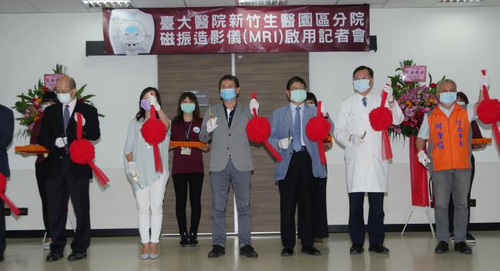
The National Taiwan University Hospital Hsinchu Biomedical Park Branch launched the 640-slice computerized tomography (CT) scanner in February. This time, an advanced magnetic resonance imaging instrument worth more than NT$70 million is launched. It will bring about more medical services and research, better disease diagnosis and improved medical quality. This morning, a press conference on the use of the magnetic resonance imaging (MRI) instrument was held. Hsinchu County Magistrate Yang Wen-Ke; Legislator Lin Wei-Chou; Chubei City Mayor Ho Kan-Ming; Chang Yun-Chong, the Director of the Department of Medical Imaging of National Taiwan University Hospital; Deputy Superintendent Chen Chang-Mu of National Taiwan University Hospital Hsinchu Branch; Lin Ming-Fong, the Director of the Medical Department and Hsiao Hsiao-Ping, the Director of the Secretary Office of the National Taiwan University Hospital Chu-Tung Branch cut the ribbon together and officially announced the launch of the magnetic resonance imaging scanner. The instrument is of the same level as that used in medical centers to provide advanced clinical diagnosis.
County Magistrate Yang expressed his gratitude to the National Taiwan University Hsinchu Biomedical Park Branch for its continuous and rapid upgrade to the latest medical equipment to provide the people in the greater Hsinchu area with better medical care and comprehensive treatment for acute and seriously ill patients. The National Taiwan University Hsinchu Biomedical Park Branch also serves as a clinical translational center. Its mission is to collaborate with manufacturers in the Biomedical Park to conduct clinical trials of new drugs or newly developed equipment. Any patients needing help can come to the hospital.
According to the National Taiwan University Hsinchu Biomedical Park Branch, coupled with the MRI scanner in the National Taiwan University Hospital Hsinchu Branch, this instrument will maximize service capacity by shortening the waiting time of patients and taking into consideration the medical needs of the local people. Preventive medicine can be actively promoted by providing comprehensive and multiple out-of-pocket health check items. People concerned about their health can freely select test items for early detection and treatment, thereby achieving preventive medicine. The hospital will also work closely with clinics and regional hospitals in the greater Hsinchu area to provide referral services and jointly care for the health of the people.
Dr. Yu, the Superintendent of National Taiwan University Hospital Hsinchu Biomedical Park said that this MRI instrument has 640 channels and uses electromagnetic vibrations to generate signals from water molecules in the body. Its ultra-high resolution water molecule diffusion imaging technology can be applied to the head, neck, entire spine, breast, abdomen, prostate and other parts of the body. The instrument not only helps with the diagnosis of neurological diseases, but can also detect any minute lesions, thereby greatly increasing the rate of cancer detection. More complicated than computer tomography, the MRI provides more accurate imaging diagnosis that will benefit people in the greater Hsinchu area. This MRI is currently the newest scanner with the highest number of channels in the Hsinchu area. Dr. Yu also mentioned that one of its characteristics is that it does not require a contrast agent to reveal blood vessel distribution and the direction of blood flow. This greatly reduces imaging time and also produces more refined images.
According to Chen Ya-Fang, Director of the Department of Imaging and Nuclear Medicine, the shape of the magnet in this new generation magnetic resonance imaging system has been optimized. It is a short magnet with a 70 cm wide hole to easily accommodate well-built patients during the examination. This model provides a wide imaging field and uniform image quality that can cover large anatomical structures and reduce examination time for patients. The shorter examination tunnel design also makes it possible to perform MRI on patients who otherwise could not receive the examination due to claustrophobia. With this advanced imaging technology, diagnostic result is also more reliable. The sound and professional scanning sequence and the latest imaging technology allow patients to receive comprehensive and radiation-free imaging information.